ANTON VAN LEEUWENHOEK (1632-1723)

1674 – Netherlands

Portrait of Leeuwenhoek

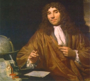
Leeuwenhoek was probably inspired to take up microscopy after seeing a copy of HOOKE’s Micrographia, though as a draper he was likely to have already been using lenses to examine cloth.
Unlike Hooke, Leeuwenhoek did not use a two lens compound microscope, but a single high quality lens, which could be described simply as a magnifying glass rather than a microscope. Leeuwenhoek is known to have made over 500 of these single–lens microscopes. They are simple devices just a few inches long, with the lens mounted in a tiny hole in a brass plate. The specimen is mounted on a point that sticks up in front of the lens. Two screws move the specimen for focusing. All else that is needed is careful lighting and a very steady, sharp eye.

After an introduction to Henry Oldenburg of the Royal Society in London from Dutch physician and anatomist Regnier de Graaf (discoverer of the egg-making follicles in the human ovary which now bear his name), Leeuwenhoek was encouraged to write to the Society’s journal ‘Philosophical Transactions’.

Leeuwenhoek’s letters were translated into Latin and English from the Dutch and he reported seeing tiny creatures in lake-water.

‘ I found floating therein divers earthly particles, and some green streaks, spirally wound serpentwise, and orderly arranged after the manner of copper or tin worms which distillers use to cool their liquors as they distil over. The whole circumference of each of these streaks was about the thickness of a hair of one’s head ’

Leeuwenhoek’s descriptions of ‘animalcules’ in water from different sources – rainwater, pond water, well water, sea water and so on – were verified by independent witnesses, including the vicar of Delft. Hooke too confirmed his findings with his own observations performed in front of expert witnesses, including Sir Christopher Wren.
Leeuwenhoek came close to understanding that bacteria were germs that cause disease but it took another century before LOUIS PASTEUR made that step.

Logo for Nikon 'Small world' competition & link to web-siteWikipedia-logo © (link to wikipedia)

NEXT buttonTIMELINE

NEXT buttonMICROSCOPY

Related sites

GERD BINNIG (GERMANY b.1947) & HEINRICH ROHRER

‘Scanning Electron Microscope’

1980 – Switzerland

SEM image of Caffeine crystals. Credit: Annie Cavanagh. Wellcome ImagesSEM image of Diatom frustule.SEM image of Moth Fly

If a needle charged with electricity is placed extremely close to the surface of a metal or semi-conductor a miniscule but measurable electric current, known as a ‘tunneling current’ will leap the gap. This current is extraordinarily sensitive to the width of the gap. The size of the tunneling current therefore reveals the distance between the needle tip and the surface.

Photograph of Gerd Binnig - worked on the scanning-tunneling electron microscope ©

GERD BINNIG

Photograph of Heinrich Rohrer - worked on the scanning-tunneling electron microscope ©

HEINRICH ROHRER

picture of the Nobel medal - link to nobelprize.org


Link to WIKIPEDIA


NEXT button - CURL_KROTO_SMALLEYTIMELINE


NEXT button - MICROSCOPYMICROSCOPY

Related sites

scanning electron microscope (serc.carlton.edu/)
how-sem-works (nanoscience.com/)
high voltage electron microscopy (esi.nagoya-u.ac.jp)(2013)

ROBERT CURL (USA b.1933) HAROLD KROTO (UK b.1939) RICHARD SMALLEY (USA 1943-2005)

1985 – USA

‘A form of the element carbon exists in which the atoms are arranged in tiny, hollow spheres shaped like soccer-balls’

Carbon may form long chains, but a structure of 60 atoms arrayed in a sphere of interlocking 20 hexagons and 12 pentagons also would form a stable structure.

Photograph of HAROLD KROTO - one of the team who first described 'Bucky balls' &copy:

HAROLD KROTO

Photograph of ROBERT CURL ©

ROBERT CURL

Photograph of RICHARD SMALLEY - one of the team who first described 'Bucky balls'

RICHARD SMALLEY

Such structures are termed ‘buckyballs’ – after architect Buckminster Fuller’s geodesic domes made from glass and metal, which demonstrate a similar structure on a macroscopic scale.

picture of the Nobel medal - link to nobelprize.org

Link to WIKIPEDIA

NEXT - ALLAN WILSONTIMELINE